A client presents his or her dog to you for squinting and redness. You apply fluorescein stain to the eye and discover a superficial ulcer. You start a topical antibiotic, pain medication and fit the dog for an E-collar. The dog returns for a recheck still squinting and the ulcer has not changed. You change medications, extend your recheck time and remind the owner to keep the E-collar on. Suddenly weeks turn into months and the ulcer will oftentimes get smaller, sometimes bigger, but always remains present and superficial. What is going on?
The cornea is made up of four layers. A superficial ulcer results from a traumatic break in the epithelium. An uncomplicated superficial ulcer should heal within 3-5 days via to cell migration and thickening. An indolent ulcer is a persistent superficial ulcer in which the epithelium has difficulty adhering to the underlying stroma due to stromal alterations. This type of ulcer is found more commonly in older dogs and Boxers.
Treatment includes aggressive epithelial debridement, which can be performed with a cotton-tipped applicator. Oftentimes this will make the ulcer larger, but only unhealthy epithelium will be removed. A dry cotton-tipped applicator works best for removing unhealthy epithelium therefore several may be used on one eye. One study showed that the addition of topical oxytetracycline (terramycin) resulted in faster healing. If these methods are not effective, and infection has been ruled out, more aggressive debridement is necessary. Further treatments include a grid keratotomy or diamond burr debridement. Diamond burr debridement is a commonly performed referral procedure for indolent ulcers. At our practice, this can be performed as an awake procedure with topical anesthetic. The ulcer is debrided first with a cotton-tipped applicator to remove loose edges. A handheld diamond-tipped burr is gently moved over the ulcer bed to remove abnormal tissue and encourage healing. Oftentimes a contact lens is placed over the corneal surface to help expedite healing. This procedure is effective 85% of the time and can be repeated if necessary. Patients continue on topical antibiotics and pain medications.
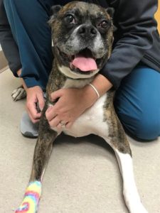
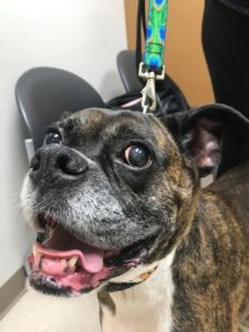
Pictured below is Prophet, a very colorful boxer who presented for evaluation of an indolent ulcer of the left eye. Oftentimes when superficial ulcers become chronic, neovascularization or granulation tissue forms across the corneal surface. This can be seen in the second picture of Prophet’s eye. The black dot over the granulation tissue is from the contact lens that was placed after the burr procedure. The last picture is Prophet at his recheck 2 weeks later. As you can see, the granulation tissue has faded and the eye is comfortable and fully healed with minimal scarring.
If diamond burr or grid keratotomy is not successful after several attempts, a superficial keratectomy can be performed in which the affected corneal tissue is surgically removed. While this procedure has an extremely high success rate, it is often reserved as a last resort for very stubborn ulcers.
Picture 1
Picture 2

Picture 3
Case submitted by: Amanda Davis, DVM, DACVO