If your pet has been diagnosed with a cataract, you likely have many questions. We’re here to help you understand your pet’s condition and how it can be treated.
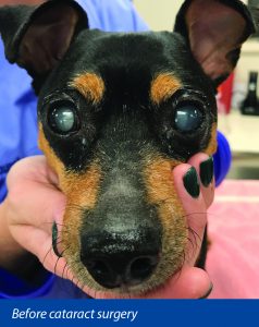
A cataract is an opacity, or cloudiness, of the lens of the eye. The lens is a clear, round structure suspended in the center of the eye that focuses light rays onto the retina. It also adjusts its shape to allow your pet to see both up close and far away. When a cataract forms, either a part of the lens or the entire lens is no longer transparent. Although light can still get through, it interferes with your pet’s vision. Looking through a cataract is often described as trying to look through a dirty window.
Cataracts can form for a number of reasons, although most are inherited. Some dog breeds are more susceptible to developing cataracts, including:
Cataracts can also form secondary to diabetes, ocular inflammation, old age, and retinal degeneration.
A cataract appears as cloudiness in the pupil (the black area in the center of your pet’s eye), but there are other conditions that can also cause cloudiness of the eye. As a normal lens ages, it hardens, which causes cloudiness that doesn’t interfere with vision. A cataract can only be differentiated from normal aging changes and other conditions through a complete ocular examination performed by a veterinarian or a veterinary ophthalmologist.
The only successful treatment available for a cataract is surgery performed under general anesthesia by a veterinary ophthalmologist. Before the procedure can be performed, further testing will determine whether your pet is a good candidate for surgery, including:
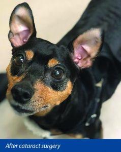
During the procedure, the lens capsule is opened, the internal lens contents are broken up and removed, and an artificial lens is typically implanted. Cataract surgery with artificial lens implantation restores normal vision approximately 90 percent of the time.
Most pets are hospitalized for one night following cataract surgery. When your pet is ready to be released, he’ll go home with an e-collar that he will wear for about three weeks to keep him from rubbing his healing eye. During recovery, your pet may be prescribed up to four different eye medications that will be dropped into the eye four times per day for several weeks.
Your pet will have several post-surgical evaluations with our veterinary ophthalmologist. Although complications from cataract surgery are rare, they do occasionally occur. Post-operative monitoring will allow us to watch for these complications:
If your pet has a cataract, schedule an evaluation with a board certified veterinary ophthalmologist. After a thorough examination, the ophthalmologist will present a treatment plan, which will likely include cataract surgery with lens implantation.
Ready to discuss cataract management for your pet or schedule an evaluation with our board-certified ophthalmologist? Contact us today.